Cochlear Endoscopy in Cochlear Implantation of a X-Linked Stapes Gusher Syndrome-Juniper Publishers
Juniper Publishers-Open Access Journal of Head Neck & Spine Surgery
Cochlear Endoscopy in Cochlear Implantation of a X-Linked Stapes Gusher Syndrome
Abstract
A 12-year-old boy with a five-year history of bilateral sensorineural hearing loss and X-linked stapes gusher syndrome developed progressive left-sided hearing loss. The pre-operative computed tomography of the temporal bone showed a bulbous internal auditory canal with a dysplastic cochlea and no apparent modiolus. A 1.3mm salivary endoscope was placed at the cochlear entrance to assess the intracochlear anatomy. This revealed membranous structures of the cochlea without direct communication to the internal auditory canal. We advocate for the use of cochlear endoscopy to better delineate inner ear anatomy, which will influence the implant selection and potentially hearing outcomes in patients.
Keywords:MCochlear endoscopy; Otoendoscopy; X-linked stapes gusher syndrome; Cochlear implant; Perilymphatic gusher.
Introduction
X-linked stapes gusher syndrome (otherwise known as X-linked deafness type 3 or Nance deafness) is a rare form of sensorineural hearing loss (SNHL) syndrome. Inherited in a sex-linked recessive manner, it is believed to be the consequence of a loss-of-function mutation on the X-chromosome in the POU3F4 gene at the DFN3 locus [1,2]. Males tend to present greater phenotypic severity than females, who present less frequently [3-5].
Affected patients have an abnormal configuration of the lamina cribrosa and internal auditory canal (IAC) [1,3,4,6,7]. This malformation leads to increased perilymphatic pressure and to stapes’ footplate fixation, giving rise to conductive hearing loss and progressive cochlear nerve incompetence. This is relevant for surgeons, as it results in an increased risk of perilymph gusher with surgical manipulation [1,3,4,6,7]. This may lead to other complications, such as otorrhea, rhinorrhea, and recurrent meningitis [8]. Meningitis complicating cochlear implantation (CI), occurs at a higher rate in patients with inner ear (IE) abnormalities, including X-linked gusher syndrome [3,9].
Traditional approaches to imaging for CI utilize computed tomography of the temporal bone (CT TB) and/or magnetic resonance imaging. We report a case whereby intra-operative otoendoscopic visualization allowed for real time visualization of the IE anatomy, which allowed us to optimize our electrode choice for CI. In this case, such visualization was valuable, as it indicated the presence of the membranous portions of the IE to evaluate if the modiolus was present, which we believe implied that a directional electrode was the most appropriate choice.
Case Report
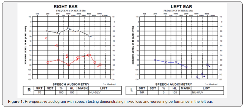
A 12-year-old boy with a five year history of bilateral SNHL and X-linked stapes gusher syndrome presented with progressively worsening left-sided hearing loss. He was initially performing well with bilateral traditional hearing aids but developed progressive mixed loss and worsening performance in his left ear. His preoperative audiogram can be seen in Figure 1. He was sent for CI candidacy assessment and was deemed to be suitable.


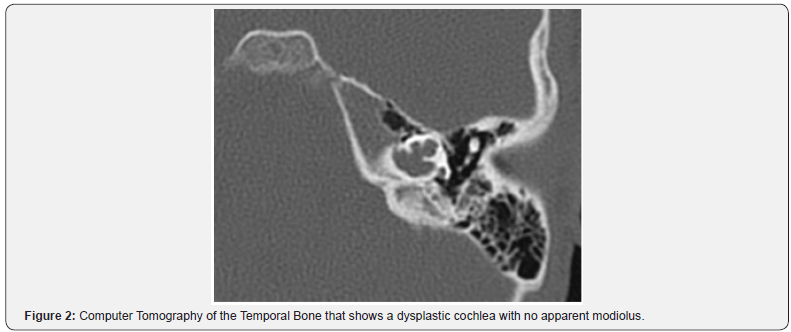
The pre-operative computed tomography (CT) of the temporal bone was assessed. This showed a bulbous IAC with a dysplastic cochlea and no apparent modiolus (Figure 2). A CI24RE (ST) implant was initially selected by the implant team due to the uncertainty regarding the location of the spiral ganglion cells.
A 1mm cochleostomy was made anteroinferior to the round window (RW). Upon entering the cochlea, a moderate perilymphatic gusher was encountered. A 1.3mm rigid salivary endoscope (Karl Storz, Germany) with a 3-chip full HD camera head (Image1 S H3-Z) was approached trans-tympannically and was placed at the edge of the 1mm cochleostomy to assess the intracochlear anatomy.
To avoid injury to the inner ear, the CSF was not suctioned. The same view was not achieved with the microscope as the endoscope provided a more magnified and higher definition view of the modiolus. There was evidence of membranous structures of the cochlea without direct communication to the IAC. Full insertion was achieved on the first attempt without any difficulty and the gusher was controlled with packing of periosteal tissue at the cochleostomy site. An intra-operative plain X-ray confirmed the electrode placement.
Discussion
When the cochlear modiolus and osseous spiral lamina are deficient, the absence of a bony septum creates a common space seen on the imaging study [10] (Figure 2); there is an abnormal communication between the IAC and the vestibule responsible for the periphymphatic gusher. Patients with IE anomalies may have both atypical positions of their spiral ganglion cells (SGC) and have a higher likelihood of having fewer spiral ganglion cells [11]. This has functional implications because at least 10,000 functional SGNs are necessary for effective speech discrimination 12]. Also this number can be further reduced by surgical trauma [12]. Therefore, choosing the proper electrode device, namely, a directional or a full banded electrode can help reduce the number of lost SGNs. The fully banded CI electrode may be more useful in the absence of a modiolus to allow for full and multi-directional stimulation, whereas the pre-curved directional electrode may be more appropriate if there is a modiolus present for precise stimulation [13].
The proper electrode choice may have important hearing outcome implications for patients with IE anomalies following CI due to the challenges of reaching an optimal level of cochlear stimulation, decreased dynamic range, a wider pulse width, and weakened neural synchrony [7]. The functionality of CI is correlated to the number of SGCs and their distances from the stimulating electrode [14].
In our patient, we assumed that there was no IAC communication due to the presence of modiolus. The moderate CSF gusher was presumed to be secondary to his X-lined stapes gusher syndrome and his enlarged IAC. Perilymph gusher during CI in patients with X-linked gusher syndrome is inevitable, but a thorough examination of the IE is still critical. CT of the temporal bone is important for surgical planning, and it is also useful to assess the likelihood of perilymphatic gusher [4]. Distinguishing features on imaging include an enlarged bulbous IAC, a widened cochlear aperture without the lamina cribrosa, cochlear hypoplasia with modiolar deficiency, and a broadening of the bony canal for the labyrinthine portion of the facial nerve [6]. Enlarged vestibular aqueducts may also appear in conjunction with modiolar deficiency [15].
The rarity of X-linked gusher syndrome may result in radiologists failing to recognize these signs, which may mislead surgeons to perform stapedectomies that are otherwise contraindicated. Thus, Incesulu et al. [3] advocated for high resolution CT of the temperal bone to assess for congenital dysplasia with ¬at least 1-mm thick slices [as opposed to 2mm thick slices], which should ideally be assessed by an experienced neuroradiologist [3]. Quan et al. [16] proposed that CT virtual endoscopy should be done to evaluate CT data through threedimensional reconstruction [16]. Limitations to both modes of assessment assume that the CT images correlate perfectly with the anatomy of the IE and that consistent radiological consultation will occur. Our case, however, demonstrated that intra-operative endoscopic findings do not always correlate with CT findings, indicating that cochlear endoscopy may be a useful tool to better delineate the intracochlear anatomy.
The otoendoscopic intracochlear view gave us accurate and real-time information on the anatomy of the IE, which confirmed the presence of a modiolus and the confidence that the CI would not be in the IAC. This has the potential to allow us to make a better-informed decision regarding the type of electrode to place.
Electrode choice is significant for patients with IE abnormalities, since the location of neural tissue may be abnormal. In patients that have an absent modiolus, a circumferentially stimulating electrode may be preferred over a full-banded electrode, which may risk adverse facial nerve stimulation [3]. One explanation for post-operative facial nerve stimulation in children with IE abnormalities is the close vicinity of the electrode to the nerve [14]. Therefore, to avoid injury, the proper choice in electrode should be made.
Conclusion
While CT temporal bone has served as the conventional approach to assessing the anatomy of the IE, fthe endoscope offers better resolution of the modiolus than the CT temporal bone, as the CT indicated that there was no modiolus. We advocate the use of intracochlear endoscopy in selected cases as it offers a better resolution than even high-resolution CT. With the potential to change electrode choices in CI, the customization of electrode choice based on the presence of membranous IE anatomy may change the hearing outcome of the patients with anomalous IE anatomy and patients with uncertain location of spiral ganglion cells.
For more articles in Open access Journal of Head Neck & Spine Surgery | Juniper Publishers please click on: https://juniperpublishers.com/jhnss/index.php

Comments
Post a Comment