Posterolateral Percutaneous Endoscopic Discectomy with Partial Pediculotomy for the L1-L2 High-Grade Downward Migrated Disc Herniation-Juniper publishers
JUNIPER PUBLISHERS-OPEN ACCESS JOURNAL OF HEAD NECK & SPINE SURGERY
Abstract
Background: Percutaneous endoscopic discectomy
(PED) is one of the most useful minimally invasive surgical techniques
for lumbar disc herniation (LDH). However, high-grade migrated disc is
difficult to treat with only the standard posterolateral approach
(posterolateral PED, PLPED).
Purpose: To overcome this difficulty, we
combined the pediculotomy with PLPED for the treatment of high-grade
migrated LDH. The pediculotomy is the recent technic for PED that has
been explored with development of high-speed drill.
Case report: A 72-year-old man had a 6-month
history of left L2 radiculopathy. His general state was too poor to
perform general anesthesia and invasive surgery. The left L1-L2 downward
migrated fragment compressed his L2 nerve root axial portion on
magnetic resonance imaging. PLPED with partial pediculotomy was used for
complete total removal, which cured his symptoms without any
complications. Thirty minutes after fragmentectomy, we needed to control
100-ml bleeding from the anterior epidural venous plexus (AEVP).
Discussion: Upper-level migrated large lumbar
disc fragments often require facetectomy with some fusion in
conventional microscopic surgery. This case was too complicated for
indicating the conventional technique. Only the flexible bipolar device
helps control hemostasis in the PED system, but some bleeding cannot be
controlled easily.
Conclusion: PLPED with partial pediculotomy
was useful for a migrated fragment in patients with poor general
conditions. Although the PED system requires a new device to control the
bleeding from the AEVP.
Keywords: Lumbar disc herniation; Posterolateral percutaneous endoscopic discectomy
Percutaneous endoscopic discectomy (PED) is one of
the most useful minimally invasive surgery for lumbar disc herniation
(LDH) (1,2). Especially, high-grade migrated fragments are difficult to
treat with the standard posterolateral PED (PLPED) technique (3,4). PED
with a high-speed drill is a minimally invasive method of approaching
the deep narrow lumbar space (5-8). This paper presents a successful
case of PLPED with partial pediculotomy for a high-grade downward
migrated fragment and discusses a hemostatic technique for severe
bleeding.
A 72-year-old man had severe untreatable lumbago and
left femoral radicular pain for 6 months. His health condition was poor
with chronic renal failure requiring dialysis 3 times per week,
hypertrophic cardiomyopathy, and obstructive ventilation impairment .The
initial physician referred the patient to us to undergo a minimally
invasive spine surgery.
During his first visit, the patient's numeric rating
scale (NRS) scores were 8/10 for low back pain and 8/10 for left front
femoral pain. His leg pain increased after 10 minutes of slow walking,
so he needed a wheelchair. Neurological examination revealed normal deep
tendon reflexes. His thigh circumference was asymmetrical (right,
35.5cm; left, 35.0cm). Owing to his pain, he could not get into a prone
position for examination with the femoral nerve stretch test. The
straight leg raising test result was negative bilaterally.
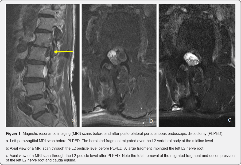
On sagittal T2-weighted magnetic resonance imaging
(MRI),a high-grade downward migrated disc was observed at the L1-L2
level on the left side (Figure 1a). The axial view at the L2 pedicle midline level showed a large fragment impinging the L2 nerve root (Figure 1b).


PLPED with partial pediculotomy under local
anesthesia was planned to decompress the left L2 nerve root. Our
anesthesiologist performed deep sedation of the patient. The patient was
placed in a prone position, and the puncture point under fluoroscopy
was simulated prior to the calculation of data (9). An 8mm skin incision
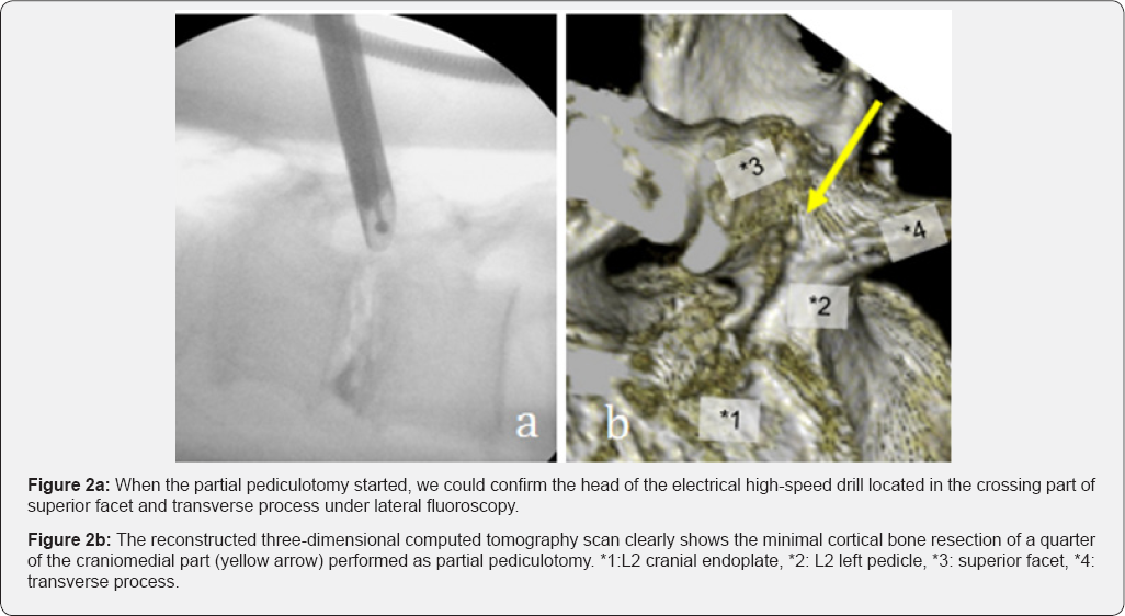
was made 5 cm to the left of the midline. The standard PLPED procedure
was started after local anesthesia. Owing to the L1-L2 foraminal severe
stenosis, we selected the outside- in technique. The adverse walking
technique could be used to approach the crossing point of the superior
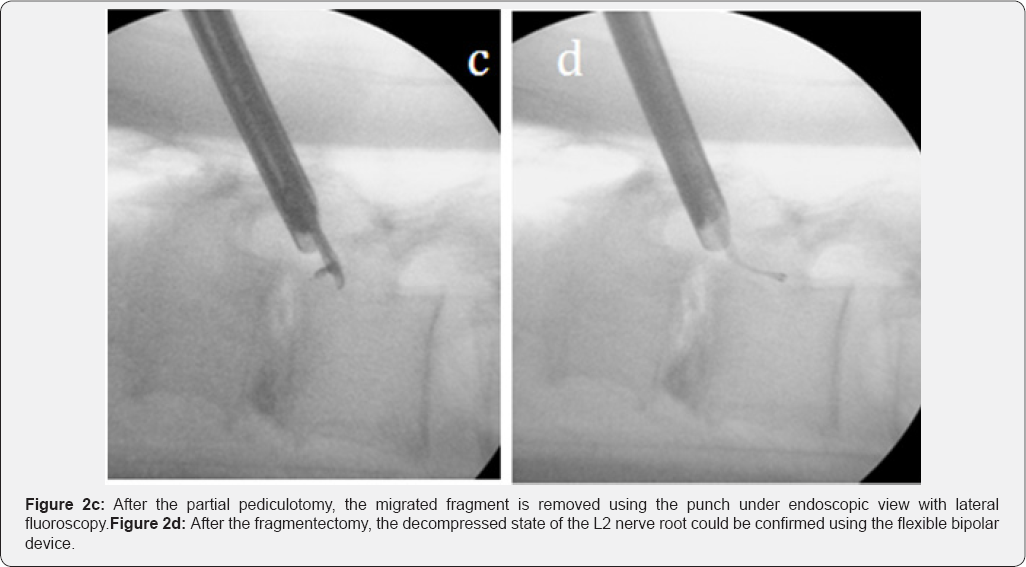
facet and transverse process (Figure 2a). Partial pediculotomy was started from this point, and a quarter of the craniomedial portion was drilled (Figure 2b) for the total fragmentectomy (Figure 2c) (10). We could decompress the L2 nerve root from the L1 endoplate level to under the midline of the L2 pedicle (Figure 2d).
The operative time was 2 hours; 100ml of bleeding
from the L1 AEVP due to injury by the tip of the Beak-type cannula took
30min to control. We first attempted the standard hemostatic techniques
of increasing the irrigation pressure (100 to 110cm H2O for 3min)
several times and compression the bleeding point with rigid dissector
but did not succeed the hemostasis. We then attempted the techniques for
such a vigorous bleeding situation include slow cannula rolling and
bleeding point compression by a flexible bipolar device with hemostatic
cotton materials. We finally achieved the hemostasis and placed a
drainage tube. Little outflow stayed only in the tube, which was
withdrawn the next morning without hematoma formation.
The patient did not have any dysesthesia and paresis.
The time to ambulation was 2 hours, and his hospital stay was 7 days.
The NRS score of his affected leg improved from 8 to 1 after 2 months.
In the next morning, his wound pain NRS score was 1. The axial view of
the MRI scan (Figure 1c) showed that the migrated fragment was completely removed.
The standard treatment for higher-level lumbar disc
herniation is conventional discectomy with some fusion after facetstomy
(11). Nowadays, many types of minimally invasive percutaneous pedicle
screw systems are available (12). Owing to the poor general condition of
our patient, we selected the operative procedures suitable for local
anesthesia were limited. Interlaminar PED (ILPED) with partial
laminectomy (7,8) and PLPED with partial pediculotomy are the best
minimally techniques for patients with poor general conditions.
The major difference between ILPED and PLPED with
partial pediculotomy is the direction of the operative approaches. ILPED
accesses from dorsal side of vertebral laminae, thereby even combined
with laminectomy the migrated fragment is still covered with neural
structures. On the other hand, PLPED with partial pediculotomy can
directly expose the migrated fragment covering neural structures. In
other words, as the migrated fragment protects underneath neural
structures, we can safely drill the inner cortical bone layer of the
pedicle. Additionally, the width of spinal canal at high vertebral level
(ex. L1-L2) is narrower than that at low vertebral level (ex. L5-S1)
(13). Consequently ILPED at high vertebral level requires more skillful
technic. Our case was affected at L1-L2 vertebral level and the migrated
fragment extended lateral part of the spinal canal (Figure 1b), we therefore planned to perform the PLPED with partial pediculotomy.
The disadvantages of PLPED under the situation of
severe foraminal stenosis include the possibility of complicating the
exiting nerve injury and bleeding from the AEVP. As the beak- type
cannula has a wider view than the square-type cannula, we changed the
square-type cannula with the beak-type cannula to complete the
decompression of the L2 nerve root. When the joystick maneuver was used
to observe the narrow disc space, the tip of the beak-type cannula
injured the L1 AEVP The flexible bipolar unit is the only instrument for
hemostasis with the PED system. We needed 30min to control 100ml of
bleeding from the AEVP The robotic general surgery system already has
many useful instruments developed for dissection and coagulation (14).
One of the urgent needs of PED surgeons is the development of a bipolar
dissector for fine manipulation like under microscopic surgery
On the basis of this case of higher-level migrated
lumbar disc herniation, PLPED with partial pediculotomy is a useful
procedure for patients with poor general conditions. For the appropriate
development of this procedure, the PED system requires new devices to
control the bleeding from the AEVP.
All the authors made a significant contribution to this case report.
To know more about Open Access Journal of
Head Neck & Spine Surgery please click on:
To know more about Open access Journals
Publishers please click on : Juniper Publishers
Comments
Post a Comment