Acute Calcific Prevertebral Tendinitis Nonvisible in Plain Radiograph-Juniper publishers
JUNIPER PUBLISHERS-OPEN ACCESS JOURNAL OF HEAD NECK & SPINE SURGERY
Abstract
Calcific tendinitis of the longus colli is an
inflammatory disease caused by calcium hydroxyapatite crystal deposition
in the longus colli tendon of the prevertebral space. The typical
imaging characteristics of this entity are calcifications on the longus
colli tendons at the C1-2 level and fluid collection in the
retropharyngeal space in plain radiograph. Usually, calcific lesion can
be detected by plain radiograph. We introduce 2 case of prevertebral
calcific tendinitis nonvisible in plain radiograph, but the diagnosis
was confirmed on computerized tomography.
Keywords: Tissue plasminogen activator; Hemorrhage; Stroke; Blepharoplasty; Ectropion Ischemic stroke; Complications; Treatment.
Many population have neck pain at some time in their
lives. According to the Global Burden of Disease 2016 Study, neck pain
is the fourth leading cause of years lost to disability, ranking behind
back pain, depression, and arthralgia [1].
And the lifetime prevalence for the rest of the adult population (18-84
years) ranged from 14.2% to 71% with a mean of 48.5% [2].
A variety of causes of neck pain have been described and include
osteoarthritis, discogenic disorders, trauma, tumors, infection,
myofascial pain syndrome, torticollis, and whiplash injury.
Unfortunately, clearly defined diagnostic criteria have not been
established for many of these entities [3]. Plain radiograph can be helpful in diagnosing neck pain.
Calcific tendinitis of the longus colli, also known
as prevertebral tendinitis or retropharyngeal calcific tendinitis, is an
uncommon benign condition caused by calcium hydroxyapatite deposition
in the longus colli tendon and inflammation of the longus colli muscle.
The clinical presentation is often nonspecific characterized by
spontaneous acute or subacute neck pain and low-grade fever. Laboratory
tests may demonstrate a mild leukocytosis and erythrocyte sedimentation
rate (ESR) is slightly elevated. Differential diagnosis between
retropharyngeal tendinitis and other diseases shows similar clinical
features, such as atlantoaxial subluxation, retropharyngeal abscess,
meningitis, infectious spondylitis. Retropharyngeal abscess is one of
the most serious disease to differentiate with calcific tendinitis.
The typical radiographic findings of retropharyngeal
calcific tendinitis are prevertebral soft tissue thickness and amorphous
calcification anterior to C1-C2. However, It is very difficult to
distinguish the early stage of retropharyngeal calcific tendinitis from
other disease, if it is invisible in plain radiograph.
We report two patients who are acute calcific
prevertebral tendinitis without any calcific mass at prevertebral area
on plain radiograph [4,5].
A 41-year-old female patient presented with a 1-week
history of a painful neck. She had no history of trauma, upper
respiratory infection. The pain gradually worsened 3 days ago from
admission. The day before admission to the hospital, the patient's pain
was aggravated worse with decreased neck motion. After hospitalization,
she complained of neck pain, stiffness of neck and dysphagia. But did
not complain fever or heating sense. On physical examination, there was
reduced active range of neck motion in all directions limited due to
pain. There was no other radiating pain or neurologic deficit to the
upper extremity. Mild tenderness was noted on the anterior and posterior
neck area. But the oropharyngeal examination was normal, with no
associated cervical lymphadenopathy. There was no abnormal findings such
as fracture, subluxation or abnormal calcification finding on cervical
spine radiograph (Figure 1).
In C-spine lateral view, the diameter of C3 level prevertebral soft
tissue thickness was 4.9mm as a normal range (C3 upper limit normal
range is 18mm) [6].
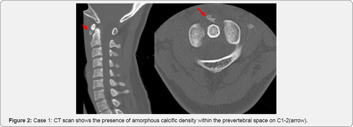
In CT and MRI, An amorphous calcification was detected, that was
8.0x6.4x3mm anterior to C1-C2 and retropharyngeal space fluid collection
was also detected at the same period of time (Figure 2).
Finally, acute calcific prevertebral tendinitis was our impression and
she was started on nonsteroidal anti-inflammatory drugs per oral.
(NSAIDS).(Aceclofenac 1 tablet, Streptokinase 1 tablet, twice a day)
After Two days on medication, visual analogue scale pain score decreased
from 7 points to 2 points.

The symptoms were improved with nearly full recovered
range of motion of neck after 10 days on medication. Laboratory tests
were slightly elevated C-reactive protein (CRP) and normal ESR. After 1
week later with treatment, ESR and CRP are decreased to the normal
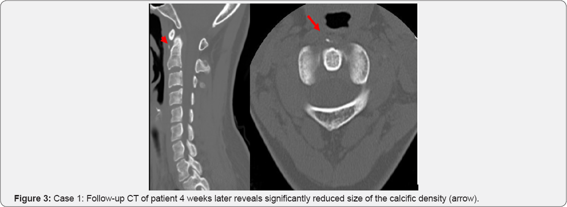
range. In follow-up CT approximately 1 month later, it was significantly
resolved calcification, size of which was 3.2x.2.1x1.0mm (Figure 3).
A 43-year-old female patient presented with one day
history of severe neck pain and aggravated by particular movements.
There was no history of fever, recent illnesses or past events. On
physical examination, we found a reduced active range of neck motion in
flexion and extension; particularly on the extension due to pain. The
rotation of the neck from side to side was a little possible. There is
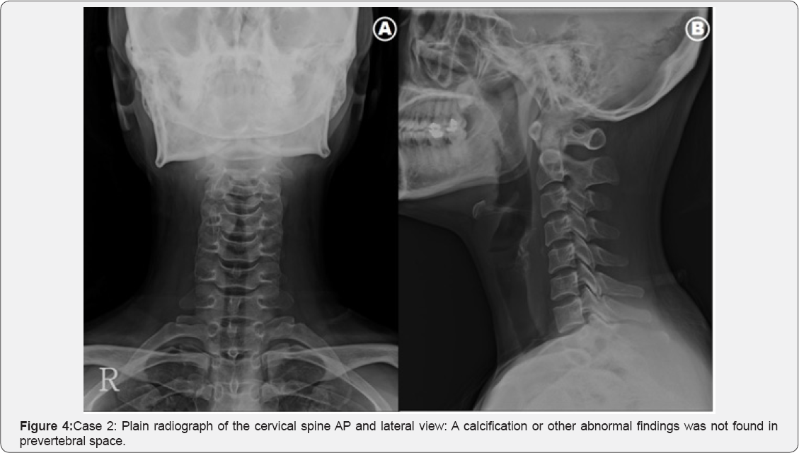
no fracture or subluxation or abnormal calcification finding on cervical
spine radiograph. In C-spine lateral view, the diameter of C3 level
prevertebral soft tissue thickness was 4.7mm and C6 level was 12.7mm; it
is within normal range (Figure 4) [6].
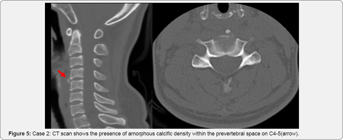
CT of the cervical spine confirmed that the calcific density mass was
irregular shape and amorphous and size was 5.3x6.3x3.5mm anterior to
C4-5 (Figure 5).
The diagnosis of acute calcific prevertebral tendinitis was made and
the patient was started on a regimen of nonsteroidal anti-inflammatory
drugs per oral. (NSAIDS). (Meloxicam 7.5mg, Eperisone 100mg, twice a
day).
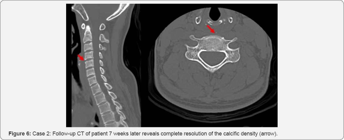
After 2 weeks, she had almost fully recovered after
receiving conservative treatment that including anti-inflammatory drugs
and rest. In 7 weeks later follow-up CT, the calcific lesion was almost
completely resolved (Figure 6).
There are many report concerning about acute calcific
tendinitis is an inflammatory disease caused by calcium hydroxyapatite
crystal deposition in the longus colli tendon of the prevertebral space.
It is also known as retropharyngeal calcific tendinitis or prevertebral
tendinitis.
Hartley first described retropharyngeal calcific
tendinitis in 1964 and was shown by Ring and colleagues in 1994 to be
due to hydroxyapatite deposition in the longus collimuscle [7,8].
The etiology of calcium hydroxyapatite crystal deposition is unclear,
and there are some suggestions that repetitive trauma, recent injury,
tissue necrosis, or ischemia may play a role.
It is believed that once the calcium crystals have
deposited within the longus colli muscle, they invoke a painful foreign
body-like inflammatory response [9].
Acute calcific retropharyngeal tendinitis affects
adults with the greatest distribution between 30 and 60 years. The
clinical pattern is nonspecific, including acute to subacute onset of
neck pain, dysphagia and low-grade fever. Dysphagia is due to the close
proximity between the retropharyngeal spaces [10].
The range of motion is extremely limited, usually secondary to severe
pain, especially in flexion and extension. Oropharynx and hypopharynx
exam performed by an otolaryngologist observed swelling and redness of
mucosa. Since WBC or CRP can be slightly elevated, lab tests do not give
significant value in differentiating calcific tendinitis from
postpharyngeal infection or abscess [11].
Retropharyngeal abscess is initially based upon clinical symptoms and
signs on physical examination. Torticollis, fever, and dysphagia
represent a classic triad of symptoms. In addition, dysphagia, and
limited neck motion [12]. Atlantoaxial subluxation presents similar symptoms with calcific tendinitis
[13].
Mainly, diagnosis is based on the clinical findings
combined with radiographic demonstration of characteristic
calcifications. Most cases show calcificated lesion in plain radiograph
and CT. The CT scan can help identify calcific deposits, which can often
appear faint on plain radiograph, if it is invisible apparent
calcification lesion on plain radiograph. The CT findings of
retropharyngeal calcific tendinitis include prevertebral soft tissue
thickness, and amorphous calcification, particularly at the anterior to
C1-C2 level [4,14]. The calcification showed low or high signal on T1 and high signal on T2-weighted MRI.
However, MRI is insensitive for the demonstration of calcification of the tendon compared to CT.
MRI is not typically necessary for this diagnosis but
can sometimes demonstrate soft tissue swelling, abnormality of disc or
vertebra, spread around in the adjacent vertebrae [14].
Retropharyngeal calcific tendinitis is a
self-limiting condition and spontaneously resolves after 1-2 weeks.
NSAIDs are usually sufficient; however, some patients may require a
short course of corticosteroids if the symptoms are severe. Most
patients can be recovered with conservative treatment such as NSAIDs in
general but difficult to considered accurate diagnosis in the absence of
radiological findings.
Acute longus colli tendinitis is a rare condition.
So, we need to distinguish more serious causes of acute neck pain such
as retropharyngeal abscess. Retropharyngeal abscess, the most feared
alternative diagnosis, can be life-threatening. It more frequently
affects young children causing fever, neck pain, dysphagia. Leukocytosis
is common. Plain radiographs often show prevertebral widening with or
without gas in the soft tissues, while CT imaging shows a hypodense
region in the retropharyngeal space with ring enhancement [15]. Atlantoaxial subluxation is also differential diagnosis to acute longus colli tendinitis and can be confirmed by CT.
Acute longus colli tendinitis is an important
consideration in the differential diagnosis of acute neck pain. If a
patient is suspected with acute longus colli tendinitis, CT should be
preferentially considered over MRI for accurate diagnosis. With the
radiographic finding, by observing that relieving symptoms with
conservative therapy such as administration of NSAlDs it will be able to
confirm the diagnosis and the treatment at the same time.
When patients present with acute symptoms such as
neck pain and dysphagia without specific findings of plain radiograph,
calcific prevertebral tendinitis should be included in the differential
diagnosis. If highly suspicious, further study with CT scan can be
helpful.
To know more about Open Access Journal of
Head Neck & Spine Surgery please click on:
To know more about Open access Journals
Publishers please click on : Juniper Publishers
To know more about juniper publishers: https://juniperpublishers.business.site/
Comments
Post a Comment