Pleomorphic Adenoma of the Tongue Base: A Case Report-Juniper Publishers
Juniper Publishers-Open Access Journal of Head Neck & Spine Surgery
Pleomorphic Adenoma of the Tongue Base: A Case Report
Abstract
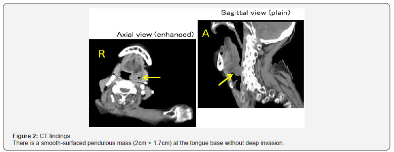
An 86-year-old woman underwent bronchoscopy after developing aspiration pneumonia. She was found to have a tumor of the tongue base and was referred to our department. Fiberscopy revealed a pendulous mass at the tongue base. On computed tomography, a smooth pendulous mass (2cm × 1.7cm) was seen at the base of the tongue, with no deep invasion. The biopsy report indicated possible mucoepidermoid carcinoma. The risk of surgery was high due to her age and co-morbidities, so the patient and her family did not agree to resection of the tumor. Aspiration pneumonia recurred several times over several months, after which she could not take anything orally and became bedridden for weeks. To improve her quality of life by minimally invasive surgery, the tumor was excised transorally under general anesthesia. The pathological diagnosis was pleomorphic adenoma, and the surgical margins were negative. The patient’s postoperative course was good. Pleomorphic adenoma often arises from the major salivary glands, especially the parotid gland, but pleomorphic adenoma of the tongue base is rare. This case is reported along with a review of the literature.
Keywords: Pleomorphic adenoma; Tongue base; Surgery
Introduction
Pleomorphic adenoma is a common benign tumor in the field of otolaryngology/head and neck surgery. It often arises from the major salivary glands, especially the parotid gland, but also from the minor salivary glands of the oral cavity. Benign and malignant tumors of the minor salivary glands are usually found on the palate, upper lip, gums, cheek, floor of the mouth, pharynx, larynx, and trachea [1]. In contrast, pleomorphic adenoma of the tongue base is rare and only 13 cases have been reported (Table 1). Here we report a patient with pleomorphic adenoma of the tongue base and review the relevant literature.

Case Report
An 86-year-old woman had noted discomfort on swallowing for several years. She developed slight dysphagia and fever six months previously. Aspiration pneumonia was diagnosed by her local physician and she was treated with antibiotics. Although her symptoms resolved within a few days, bronchoscopy revealed a tumor at the tongue base and she was referred to our department. Transnasal fiberscopy demonstrated a pendulous mass at the tongue base (Figure 1). Computed tomography revealed a smooth-surfaced pendulous mass (2cm × 1.7cm) at the tongue base without deep invasion (Figure 2). Biopsy of the tumor gave a diagnosis of possible mucoepidermoid carcinoma. It was considered that this tumor might have caused her dysphagia and aspiration pneumonia. She had a history of diabetes mellitus, schizophrenia, femoral fracture, and dementia.

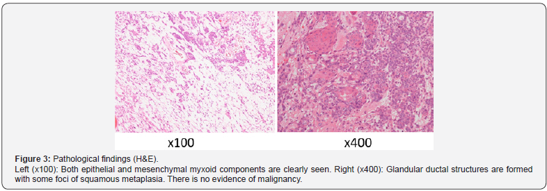
Surgery was considered to be high risk due to her age and co-morbidities. Because the patient and her family did not agree to resection of the tumor, she was followed up by her local physician. Aspiration pneumonia recurred several times over several months, after which she could not take anything orally and became bedridden for weeks. To improve her quality of life by minimally invasive surgery, the tumor of her tongue base was excised transorally under general anesthesia. The working space in the oral cavity and pharynx is limited, so we resected the mass by using laparoscopic instruments. The postoperative pathological diagnosis was pleomorphic adenoma and the surgical margins were negative (Figure 3). After surgery, she could eat without discomfort on swallowing or recurrence of aspiration pneumonia. The tumor has not recurred after followup for seven months (Figure 4).

Discussion
Pleomorphic adenoma was first described by Missen in 1874. About 80% of pleomorphic adenomas arise in the parotid gland, followed by 10% in the submandibular gland and 10% in the minor salivary glands [2]. Tumors of the minor salivary glands usually arise on the palate, upper lip, gums, cheek, floor of the mouth, pharynx, and trachea [1]. The most frequent site for pleomorphic adenomas of minor salivary glands is the palate (50%), followed by the upper lip [3]. In contrast, pleomorphic adenoma rarely arises from the tongue base and only 13 cases have been reported previously (Table 1) [2,4-15].
Surgery is the accepted treatment for pleomorphic adenoma and the tumor was subjected to surgical resection in all of the previous reported cases. Because of its anatomical features, approaching the tongue base for surgery raises several problems. In particular, the site is difficult to view by direct vision and the working space is narrow. The surgical approach depends on the size and location of the tumor, so the surgeon should plan treatment carefully. Various surgical approaches have been used, including the transoral, transhyoid, transpharyngeal, transmandibular, and combined transoral-transcervical approaches. We performed transoral excision to minimize surgical invasion, because the patient was elderly and had a history of schizophrenia and dementia, suggesting that brief hospitalization was required. The tumor was pedunculated and not deeply infiltrative, so we decided that transoral resection was reasonable. Because the working space in the oral cavity and pharynx is very narrow, laparoscopic instruments were used. However, the devices were actually too long for the transoral approach, so a new approach such as robot support is needed for resection of tongue base tumors [16].
For more articles in Open access Journal of Head Neck &
Spine Surgery | please click on: https://juniperpublishers.com/jhnss/index.php
For more about Juniper Publishers | please click on: https://juniperpublishers.com/pdf/Peer-Review-System.pdf

Comments
Post a Comment