Delayed Progressive Cauda Equina Syndrome after Transverse Sacral Fracture: A Case Report-Juniper Publishers
Juniper Publishers-Open Access Journal of Head Neck & Spine Surgery
Delayed Progressive Cauda Equina Syndrome after Transverse Sacral Fracture: A Case Report
Abstract
Transverse sacral fractures are infrequent injuries that often cause neurological impairment referred to as cauda equine syndrome. To the best of our knowledge, the onset of neurological symptoms can be delayed up to 2 months. We report a case of isolated transverse sacral fracture through S2-S3 which caused minor symptoms after 6 days. Major neurological disability appeared with a 3 months delay. A limited laminectomy was performed with good clinical results. The 3 months delay appeared to be the longest compared to available literature.
Keywords:Transverse sacral fracture; Laminectomy; Caudaequina syndrome; Delay
Introduction
Transverse sacral fractures are rare. They occur after low or high energy trauma, with clinical manifestations that include neurological dysfunctions. According to the literature, these symptoms can be delayed [1-4]. To our knowledge, the literature reports only 2 cases with neurological signs that became significant as late as 2 months after trauma [1]. There is no clear consensus on how to manage these fractures [4-6]. We report a case of isolated transverse sacral fracture through S2-S3 which caused significant neurological symptoms 3 months after trauma. We compare our observation to the available literature.
Case Report
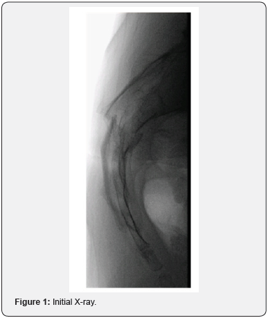
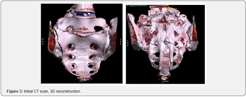
A 50-year-old woman presented to our emergency department with pain in lumbo-sacral region two days after falling in the stairs (approximately 3 meters) with impact on her lower back. She had no relevant medical history. The pain increased in seated and supine position. Bowel, bladder, sphincters and lower limbs were clear of any symptom. Perineal sensation was normal. X-rays and CT scan images revealed a transverse sacral fracture through S2-S3 junction with no vertical extension. The distal fragment was flexed and anteriorly displaced of 5mm. The canal was enchroached by fragments from the anterior and posterior aspect of the sacral canal (Figure 1-3). She was discharged with painkillers and followed in outpatient clinic.


At day 6 after trauma, she had an additional symptom of saddle hypo-esthesia. Bladder, sphincters and lower limbs were still normal. The conservative management was continued and surgical treatment was to be considered only in case of neurological deterioration. At 6 weeks, pain had decreased but saddle numbness persisted with no aggravation nor additional symptom.
Urinary incontinence appeared at 3 months post trauma and was complete within 2 weeks. CT scan imaging was repeated and showed advanced consolidation with a callus that was slightly larger than on previous workups. This suggested increased canal compression (Figure 4). The patient underwent S2-S3 laminectomy. Urinary dysfunction was almost back to normal on the day after surgery. All neurological symptoms recovered within two weeks following surgery.

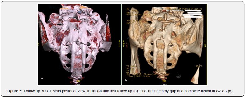
At last follow up (12 months after initial trauma), the patient’s neurological status was normal. She had resumed regular jogging and swimming with no discomfort. Final CT scan showed full fusion of the fracture and post laminectomy status in S2-S3 (Figure 5).

Discussion
Several classifications have been proposed for sacral fractures but none of them encompasses all the fractures’ patterns encountered in clinical practice. Roy-Camille et al. [7] described suicidal jumper’s fractures that occurred at S1-S2 level comprising transverse and vertical orientations (H, U patterns). Their study classified 3 types of fractures.
a) Type 1: Anterior flexion fracture.
b) Type 2: Anterior flexion fracture with posterior displacement of proximal fragment.
c) Type 3: Extension fracture with anterior displacement of proximal fragment.
Denis et al. [8] classified sacral fractures in 3 vertical zones: zone 1 lateral to the foramina, zone 2 involving the foramina and zone 3 the sacral canal [4,5,8]. Zone 3 lesions can be subclassified into vertical or transverse fractures [5].
Shmidek et al. [9] divided transverse sacral fractures according to the level: high (through S1-S2) versus low (through S3-S5) [5,9]. High fractures are caused by indirect high energy forces (motor vehicle accident, suicidal jump) and are usually unstable due to a three-dimensional configuration (H,U or Ttypes), whereas low fractures result from direct trauma (fall onto buttocks) and are likely to be stable [2,5]. Given the sacroiliac weight transmission, these fractures are stable if sacrum and sacroiliac joint above S1 foramen are intact [5].
Our patient fell onto her buttocks and presented an isolated transverse fracture through S2-S3 that can be classified as a low transverse Zone 1-2-3 fracture. It has been observed that neurologic injury is most frequent in fractures involving Denis Zone 3 (60%) [4,5,10]. Denis et al. [8] found the risk to be greater in transverse fractures than in vertical ones [2,8]. Usual clinical repercussions are bowel and bladder dysfunction, sphincters incontinence, L5/S1 deficits and saddle anaesthesia resulting in cauda equina syndrome [2,4,5]. According to the literature, these neurological signs are often variable and delayed [1,3], while local pain is always noted from the beginning [11]. The delays for neurological symptoms range from a few days to 2 months as reported by Lee et al. [1], with most neurologic deficits appearing at the same time. Aresti et al. [4] report a case with progressive symptoms starting with isolated S1 radicular symptoms at day 10 after trauma followed by urinary dysfunction and saddle anaesthesia 6 weeks later. In the present case, saddle hypoesthesia appeared at day 6 and remained stable until urinary incontinence appeared at 3 months post trauma.
After physical examination, transverse sacral fractures can be confirmed and evaluated on plain X-rays. However, CT scan imaging is the best way to avoid misdiagnosis and allow for thorough description and classification [2,11].
The literature does not provide precise treatment guidelines. In the case of transverse sacral fracture with neurological impairment, there is controversy between surgical and conservative management. Both approaches do result in recovery rates as high as 80% [6]. Surgery has been advocated in case of high energy trauma, neurologic deficit, displacement exceeding 1cm, canal encroachment and fracture instability [2,5,6]. Timing of surgery is still debated and there’s no clear evidence whether early or late intervention is best [2,4]. As to the surgical technique, decompression alone by wide laminectomy has been proposed in case of neurological impairment with canal stenosis [1,2,4-6]. Internal fixation is recommended in case of instability or major displacement [2,4,6].
In the present case, the early neurologic impairment was considered as moderate and priority was given to conservative treatment. Surgery was indicated by the appearance of incontinence. Laminectomy was performed considering the canal encroachment while stabilization was unnecessary: fusion was already obtained, especially in the anterior column. Anatomical and clinical results were satisfying at final follow up.
Conclusion
Isolated transverse sacral fracture is rare and likely to occur after low energy direct trauma. Clinical manifestations include local pain and cauda equine signs that are variable in time and severity. Mild neurological deficits can deteriorate even 3 months later. Management should be adapted to each patient, according to the fracture pattern and neurological impairment. Sacral decompression can still result in good improvement, even at a delayed state.
For more articles in Open access Journal of Head Neck & Spine Surgery | Juniper Publishers please click on: https://juniperpublishers.com/jhnss/index.php

Comments
Post a Comment