A Rare Case of Traumatic Leptomeningeal Cyst in Adult: Case Report-Juniper publishers
JUNIPER PUBLISHERS-OPEN ACCESS JOURNAL OF HEAD NECK & SPINE SURGERY
Abstract
Roux-en-Y choledochojejunostomy is a common bile duct
reconstruction operation, which has a good surgical effect, and has a
low chance of bile leakage and bile duct stenosis. The main
complications are bleeding, infection, anastomotic leakage and stress
ulcer, but unclosed mesenteric foramen of small intestine after
Roux-en-Y choledochojejunostomy is rare. Therefore, in this report, we
reviewed a case of the clinical data, imaging findings, and surgical
status of a patient with unclosed mesenteric foramen of small intestine
of ten years after Roux-en-Y choledochojejunostomy and hope to provide a
case reference for clinicians.
Keywords: Roux-en-Y choledochojejunostomy Unclosed mesenteric foramen Surgery suture
Introduction
Roux-en-Y choledochojejunostomy has different
complications in clinical [1,2], and it is rare to have a proximal
unclosed mesenteric hiatus. In this case, the patient had an
intermittent colic episode, which was considered as the possibility of
intestinal volvulus by CT examination. The small intestinal volvulus was
diagnosed by postoperative exploration, and the unclosed mesenteric
hiatus of the small intestine was also found. Therefore, through this
case report, it is hope that can help clinical diagnosis and treatment
in this area.
General clinical data and CT findings of this patient
A 51-year-old female patient, recently the lower
abdomen is painful for 2 days, and was admitted to the hospital for half
a day with increased pain. This patient had a history of open
choledochectomy, and Roux-en-Y choledochojejunostomy for more than 10
years. Examination revealed that the patient’s abdomen was soft,
tenderness in the lower abdomen, no obvious rebound pain, and active
bowel sounds. T: 36. 8℃, R: 19 /min, P: 78 /min, BP: 130 /80 mmHg.
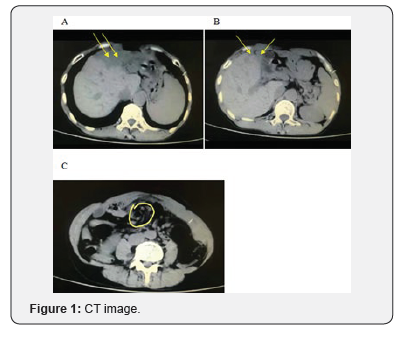
Abdominal CT examination showed that multiple dilatation of the
intrahepatic bile duct, a large low-density shadow and a lower-density
shadow could be seen in the left lobe of the liver, the position of the
gallbladder fossa not shown the gallbladder, the density of the
surrounding tissue was vague, and a small amount of free gas could be
seen, the proximal small intestine stenosis and the thickened peritoneal
adhesions could be seen (Figure 1). The image of intestinalvolvulus and
mesenteric hiatus not closed.after Roux-en-Y choledochojejunostomy. A
and B: Lamellar low-density shadows and the lower liquid density shadows
can be seen in the left lobe of the liver, surrounding tissues are
blurred, gas density shadows can also be seen, and dilatation of the
intrahepatic and external bile ducts can be seen. C: Circular stenosis
can be seen in the upper segment of the small intestine the density of
intestinal wall is uniform. These images suggest that it may be
intestinal volvulus, but the mesenteric hiatus cannot be found.

Operation situation
The patient was anesthetized and placed on the operating
table, and then routinely the abdomen was disinfected, and the
sterile sheet was paved. An incision was made in the midline of
the abdomen, and then the abdominal wall, rectus abdominis, and
rectus sheath was opened in order, and entered the abdominal
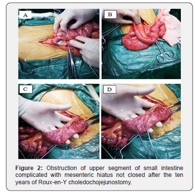
cavity. The bowel was detected, it was found that the bowel at
the proximal anastomosis of the upper segment of jejunum was
twisted, resulted in intestinal stenosis and intestinal wall edema,
and further exploration revealed that the unclosed mesenteric
hiatus near the site of intestinal stenosis (Figure 2). After the
reduction of intestinal volvulus, the mesenteric hiatus was
sutured and closed, and then further detected whether there
are any abnormalities in other intestines. Subsequently, the
abdominal incision was sutured, and the wound was covered
with sterile gauze. The bleeding and anastomotic leakage were
not found during the operation. The map of obstruction of upper
segment of small intestine complicated with mesenteric hiatus
not closed. A: The site of volvulus of the small intestine, located
the below anastomosis and the upper segment of the jejunum. B:
The site of intestinal volvulus has been restored and shows the
site of choledochojejunostomy. C and D: The site of the proximal
mesenteric hiatus of the small intestine has shown and the
mesenteric foramen is closed by stitching.

Discussion
Roux-en-Y choledochojejunostomy is to cut off and close
the distal end of the jejunum about 15 cm from the duodenum
jejunum, retain the broken suture for traction, and jejunumjejunostomy
is performed at a distance of 55 cm from the
jejunum [3]. It is one of the commonly used surgical methods in
general surgery []. The main indications are as follows [4-6]:
a) Benign stricture of bile duct.
b) Some diseases requiring extrahepatic bile duct
reconstruction (such as, choledochal cyst, choledochal
malignant tumor or pancreatectomy, etc).
c) Liver transplantation is not suitable for end-to-end
biliary anastomosis.
d) Common bile duct stricture caused by trauma, surgery
or malignancy.
e) Distal common bile duct obstruction caused by
malignant tumors of pancreas, duodenum and bile duct,
and incarcerated stones. Roux-en-Y choledochojejunostomy
has become an important surgical method for the treatment
of biliary diseases, which can significantly improve biliary
obstruction and relieve symptoms [7].
However, there are certain contraindications, mainly those
with intrahepatic stenosis or stones above the common bile duct
that have not been treated [8]. The long-term complications
of the Roux-en-Y choledochojejunostomy are mainly bleeding,
infection, anastomosis and leakage, and stress ulcers [9,10].
The main cause of postoperative bleeding is the peeling surface
bleeding and the instability of vascular ligation during operation,
while the coagulation factor or fibrinogen deficiency is also part
of the cause of bleeding [11]. The principle for the treatment of
postoperative bleeding is through blood transfusion, transfusion
and correction of blood coagulation. If the vital signs can remain
stable, the patients can continue to be treated conservatively,
however, if the vital signs are unstable, it is necessary to reexplore
in time to stop bleeding [12]. Patients can be cured by
suing antibiotics after simple infection however, retrograde
biliary infections are often secondary to anastomotic stenosis
and stone formation [13]. The main causes of anastomotic
stenosis and biliary fistula are the high position of the resected
bile duct and small anastomosis diameter. Poor technique during
operation, ischemia of the bile duct wall and scar contraction
can also lead to anastomotic stenosis, which can further lead to
poor bile drainage and intestinal reflux, cause reflux cholangitis
and aggravate symptoms. Large caliber choledochojejunostomy
or the use of a silicone tube to support the drainage tube can
effectively prevent the development of the postoperative
anastomosis stenosis and biliary fistula [14,15]. In this report,
we reviewed a patient with Roux-en-Y choledochojejunostomy,
due to this patient has a long postoperative time, we only
considered the possibility of small bowel torsion before surgery.
Fortunately, the location of the unclosed mesenteric hiatus in the
small intestine was close to the intestinal torsion, so it was found
during surgery. If neglected, serious consequences were brought
to the patient after surgery. Therefore, we reported the clinical
symptoms, CT imaging manifestations and surgical conditions of
the patients accordingly, and as a reference for clinicians.
To know more about Open Access Journal of
Head Neck & Spine Surgery please click on:
To know more about Open access Journals
Publishers please click on : Juniper Publishers
Comments
Post a Comment