Unilateral Bleeding Polyp in a Child: Lobular Capillary Hemangioma-Juniper publishers
JUNIPER PUBLISHERS-OPEN ACCESS JOURNAL OF HEAD NECK & SPINE SURGERY
Abstract
Epistaxis, though being common, always attracts
special attention from medical professionals due to the nature of
bleeding involved in the process. Children presenting with unilateral
epistaxis warn otolaryngologists as the cause can vary from nasal
foreign bodies to life threatening hemangiomas and angiofibromas. We
present a rare case of lobular capillary hemangioma originating from
nasal septum in a child who presented with episodes of profuse bleed.
Early diagnosis and management with total excision was awarding. The
occurrence is rare, however it should be in the differential diagnoses
of unilateral nasal bleed in outpatient and emergency settings and
strong suspicion is warranted.
Introduction
Lobular capillary hemangioma (LCH) is an acquired
benign vascular proliferation of unknown origin, which was first
described as human botromycosis by Poncet and Dor in 1897 [1].
Skin of head and neck and mucos membrane of the oral cavity are common
sites but LCH is rare in the nasal cavity of children. The nasal septum
is the most common affected nasal site, vestibule and middle turbinate
being the other involved sites [2,3]. We present a case of lobular capillary hemangioma in a nine year old child who had presented with unilateral nasal bleed.
Case Report
A nine year old child presented to the outpatient
department of otolaryngology and head and neck surgery with 5 months
history of recurrent nasal bleeding from left nasal cavity and left
sided nasal obstruction. History of frequent nose picking present. On
anterior rhinoscopy, single red fleshy lobulated mass of size 1.5x1.5cm
was present in the left nasal cavity surrounded with blood clot.
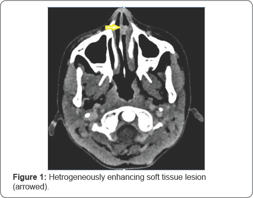
Contrast enhanced Computed tomography (CECT) of Paranasal sinus revealed
a heterogeneously enhancing soft tissue lesion attached to the
cartilaginous part of nasal septum (Figure 1).
Rest of the head and neck and systemic examination was normal. Hence
provisional diagnosis of a vascular nasal mass was made. Patient was
planned for endoscopic excision under general anesthesia.

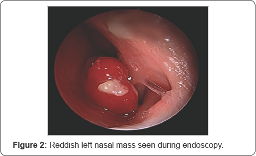
Under general anaesthesia, after decongestion of
bilateral nasal cavity, evaluation of the nasal mass was done.
Endoscopic evaluation revealed a pinkish nasal mass which was arising
from the cartilaginous part of the nasal septum (Figure 2).
Stalk of the nasal mass was cauterized with bipolar cautery and the
tumour was removed into to with sacrifice of 0.5cm surrounding mucosa at
the site of attachment of the lesion. Adequate haemostasis was
achieved. Anterior nasal packing was done. Anterior nasal packs were
removed after 24 hours and patient was discharged. The follow up of the
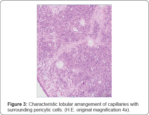
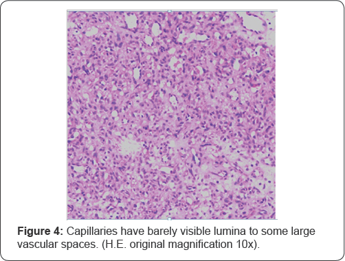
patient was uneventful, Histopathological examination revealed the
diagnosis of lobular capillary hemangioma. Microscopic examination
showed biopsy tissue lined by focally ulcerated stratified squamous
epithelium with underlying lobular arrangement of capillaries
surrounding a large central vessel. The lumen of these capillaries
varied from barely visible to large vascular spaces. The spindle shaped
pericytic cells were seen in the periphery. No atypical mitotic figure
or evidence of necrosis (Figure 3 & 4).


Discussion
Lobular capillary hemangioma (LCH) was thought as
pyogenic granuloma in earlier times but later denoted as misnomer
because of non-infectious and non granulomatous nature. Present
terminology of LCH is based on the characteristic histopathological
findings described by Miller [4].
Incidence of LCH is more in females as compared to males and is common in third decade of life [5].
Nasal cavity is an unusual site for LCH. Gingiva is a common site of
lobular capillary hemangioma. Among the nasal sites, anterior septal
mucosa and tip of turbinate's are the most frequent involved sites [1].
The presented case is that of a 9 year old male child who presented
with a bleeding polypus of the anterior nasal cavity and which turned
out to be LCH. The lesion in this case was found to be attached to the
septal cartilage.
Natural history and progression of LCH involves
many contributing factors. These include trauma, hormonal imbalances,
viral oncogenes, arterioveno malformation and angiogenic growth factors
etc [6].
This child had a history of frequent nasal pricking, which could have
led to repeated microtrauma and proliferation of vessels to form LCH
attached to the nasal septum anteriorly. LCH have also been described as
pregnancy tumour [7] due to increased associations with high levels of estrogen and progesterone during pregnancy.
LCH usually presents with various symptoms such as
unilateral epistaxis, nasal obstruction, nasal discharge, facial pain,
headache etc depending on the site of lesion [6].
This patient had presented to our OPD with repeated episodes of left
sided nasal bleed especially after nose pricking. Father of the child
had noticed a small red mass in the left nasal cavity for which he
brought the child into our outpatient department.
CECT of the nose and paranasal sinuses is the
preferred investigation for diagnosis of vascular lesions. Magnetic
Resonance Imaging (MRI) with T2 weighted images reveal vascular soft
tissue lesions with multiple flow voids [8].
Criterion standard treatment in LCH has always
been complete surgical excision of the lesion. Surgical technique and
instrumentation used for such kind of lesions include cold
instrumentation (with or without preoperative embolization),
electrocoagulation, cryotherapy and LASER [9].
We did endoscopic excision of the hemangioma followed by cauterization
of its base at the nasal septum which had the feeding vessels in it.
Recurrences of the lobular capillary hemangioma
are a rare occurrence. However, evidence in literature report a
recurrence rate of 0 to 42% depending on the location, extent of removal
and time of follow up [10]. This case has been followed up for 4 months till date and there is no evidence of recurrence of the lesion.
Conclusion
Lobular capillary hemangioma of the nasal cavity
is a rare entity especially in a male child in first decade of life.
However, it should be considered as one of the differential diagnoses of
unilateral epistaxis in children. High index of suspicion is the key
for identifying such a lesion. Endoscopy guided complete surgical
excision of the lesion is the management of choice for nasal lesions.
Recurrence is rare with no malignant transformation reported till date.
To know more about Open Access Journal of
Head Neck & Spine Surgery please click on:
To know more about Open access Journals
Publishers please click on : Juniper Publishers
Comments
Post a Comment