Incidence and Prognostic Factors in Distant Metastasis from Primary Head and Neck Cancer-An Institutional Experience-Juniper publishers
JUNIPER PUBLISHERS-OPEN ACCESS JOURNAL OF HEAD NECK & SPINE SURGERY
Abstract
Introduction: Distant metastases adversely
affect treatment planning as well as the overall prognosis of patient.
The incidence of distant metastases is less in primary head and neck
malignancy than other malignancies. objective of to calculate the median
interval and to identify the frequency of occurrence as well as various
risk factors for distant metastases in patients with head and neck
squamous cell carcinomas.
Materials and Methods: Retrospective data of
500 patients operated at HCG Manavata cancer centre, Nashik for head and
neck cancer was obtained from hospital management software system from
year 2010 to 2014 with follow up of at least 2.5 year. The following
variables were evaluated in relation to distant metastases: age, gender,
TNM stage (I-IV), histological grade, and the site of origin of the
tumor.
Results: Male to female ratio was 4.2:1,
Distribution of primary site constituted buccal mucosa (25%), tongue
(50%), lower alveolus (25%). Overall incidence of distant metastases in
head and neck cancer was 3.2%.
Conclusion: Patients with advanced stage
disease, higher histological grade as well as with extracapsular
extension should be kept under close observation with periodic
screening.
Keywords: Distant; Metastasis; Prognosis; Advanced stage Introduction
In spite of tremendous progress in radiotherapy and
chemotherapy in the past 3 decades, overall survival of head and neck
cancer patient has not been significantly improved [1,2].
Distant metastases adversely affect treatment planning as well as the
overall prognosis of patient. The incidence of distant metastases is
less in primary head and neck malignancy than other malignancies.
Location of primary tumor and clinical TNM staging significantly affects
the incidence of distant metastasis and it is also influenced by the
presence or absence of regional control above the clavicle. Primary
tumors of advanced T stages in the hypopharynx, oropharynx and oral
cavity are associated with the highest incidence of distant metastases.
Distant metastasis is commonly seen in lung region accounting for 66%.
It may be difficult to distinguish pulmonary metastasis from a new
primary tumor, particularly if solitary. Other metastatic regions
include bone (22%), liver (10%), skin, mediastinum and bone marrow. This
retrospective study was conducted at HCG Manavata Cancer Centre, Nashik
with the objective to calculate the median interval and to identify the
frequency of occurrence as well as various risk factors for distant
metastases in patients with head and neck squamous cell carcinomas.
Materials and Methods
Retrospective data of 500 patients operated at HCG
Manavata cancer centre, Nashik for head and neck cancer was obtained
from hospital management software system from year 2010 to 2014 with
follow up of at least 2.5 year.
Inclusion criteria
a) Age 20 to 80 years of patient.
b) Patient with primary head and neck malignancy.
c) Patient with stage I, II, III, IV disease.
d) Both male and female sexes.
The following variables were evaluated in relation to
distant metastases: age, gender, TNM stage (I-IV), histological grade,
and the site of origin of the tumor. After treatment patients were
followed up every month for atleast 1 year with regular screening for
lung metastasis with chest x-ray and ultrasonography for any
locoregional recurrence, after one year of regular follow up patient
were recalled after every 3 months with routine investigation to rule
out any distant metastasis. At our institute patients with advance stage
disease has been put under close observation with yearly PET CT scan to
rule out any occult distant metastasis.
Results

Male to female ratio in recurrent cases was 4.2:1 (Figure 1).
Patients with primary tongue malignancy showed aggressive behaviour
even in Stage I disease. The median interval was measured between
confirmation of diagnosis to the occurrence of distant metastases.
Average median interval with tongue primary and stage I disease was only
3 months. Our institutional experience of incidence of distant
metastasis was accounting for 3.2% of cases.
In our study skeletal metastasis was observed in 2 5%
of patients among all the distant metastasis. Patients with skeletal
metastasis were mostly presented with stage III and stage IV disease
while one patient was seen with stage I disease. 50% of the patients
were having tongue as primary lesion while 25% of the patients were
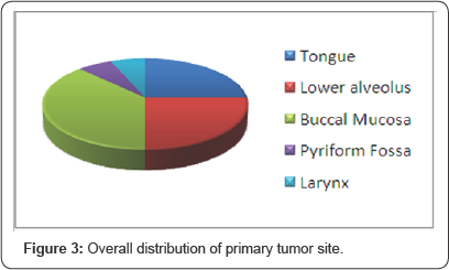
presented with buccal mucosa and lower alveolus as the primary each (Figure 2).
Patients with multiple bony metastases were 25% with involvement of
vertebrae, scapula, tibia and maxilla as compared to solitary metastasis
which was found to be 75%. In our study extracapsular spread was
observed in 75% of patients with skeletal metastasis, average median
interval was observed to be 6.25 months. In this study skeletal
metastasis is exclusively seen in male with 100% incidence. In our study
distant failure rate was observed 3.2% out of which cutaneous
metastasis was observed to be 31.25% which is quite high as compared to
other study. Patients with cutaneous metastasis were presented with
stage III and stage IV disease with one patient was observed to be a
stage I disease and tongue primary. Average median interval for
occurrence of distant metastasis was observed to be 118 months.
Histological grading shows that patient with grade III diseases
comprises of 40% compared to grade II which is 20% and grade I
constituting 40% of cases. In our study liver metastasis was very rare
and found in only one patient with rate of 6.25% of cases. In this
patient median interval was 36 months which is quite good compared to
other studies (Figure 3).


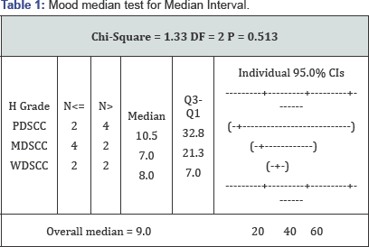
Mood's Median Test was used to derive relationship
between histological grade and median interval. It showed that average
median interval in PDSCC is 10.5 months; in moderately differentiated
squamous cell carcinoma (MDSCC) it is 7 month whereas in well
differentiated squamous cell carcinoma (WDSCC) it is 8 months. Variation
of median interval in poorly differentiated squamous cell carcinoma
(PDSCC) is comparatively larger as compare to the variation in other two
groups. Statistically average median interval in all three category
patients is nearby same (Table 1).
Table 1
show that average median interval in PDSCC is 10.5 months; in MDSCC it
is 7 month whereas in WDSCC it is 8 months. Variation of median interval
in PDSCC is comparatively larger as compare to the variation in other
two groups. Statistically average median interval in all three category
patients is nearby same.
Discussion
Squamous cell carcinoma of head and neck region
commonly spread through the lymphatic spread while the non lymphatic
spread accounts for approximately 10% of cases involving lungs, brain,
bones and skin [3].
Risk factors for head and neck cancer metastasis have been already
studied and confirmed that the increased risk of metastasis is
associated with stage of disease, its histological grade, size of
primary lesion and its site of occurrence with hypopharynx accounting
60%, base of tongue 53%, anterior tongue 50% [4]. Distant metastasis in head and neck cancer patient ranges from 6% to 43% in autopsy cases [2,5]. While according to Betka's review in his clinical studies it accounts for 8%-17% [6]. While in our study conducted at our institute the incidence of distant metastasis is quite low accounting for 3.2% of cases.
Skeletal metastasis
Squamous cell carcinoma of head and neck region
predominantly metastasizes to lung which constitutes about 66% of
distant metastases. Solitary pulmonary metastasis is difficult to
distinguish from a new primary tumor SCC of head and neck region also
metastasizes to bone (22%), liver (10%), skin, mediastinum and bone
marrow [7].
In our study skeletal metastasis was observed in 25% of patients among
all the distant metastasis. Patients with skeletal metastasis were
mostly presented with stage III and stage IV disease while one patient
was seen with stage I disease. In this study 50% of the patients were
having tongue as primary lesion while 25% of the patients were presented
with buccal mucosa and lower alveolus as the primary lesion each.
Patient with multiple bony metastasis were 25% with involvement of
vertebrae, scapula, tibia and maxilla as compared to solitary metastasis
which was found to be 75%. In our study extracapsular spread was
observed in 75% of patients with skeletal metastasis, which denotes
extracapsular spread is a crucial factor in bone metastasis.
Histological grade shows that patient with moderately differentiated
squamous cell carcinoma (MDSCC) accounts for 75% of bone metastasis
compared to well differentiated squamous cell carcinoma (WDSCC) with 25%
of cases. Patients with skeletal metastasis has poor prognosis with
disease free survival of only few months [8].
In our study average median interval was observed to be 6.25 months.
Patients with multiple bone metastasis was having less median interval
as compared to solitary metastasis. In this study skeletal metastasis is
predominantly seen in male with 100% incidence in male patients.
Skin metastasis
Squamous cell carcinoma of head and neck region
rarely metastasise to skin and associated with poor prognosis and
progressive disease [9]. Incidence of distant cutaneous metastasis is in the range of 0.7% to 10% and is seen in breast and lung as primary tumor [10].
Incidence of cutaneous metastasis from squamous cell carcinoma of head
and neck region is lower as compared to other primary malignancy and
accounts for 0.8-1.3% [11].
In our study distant failure rate was observed 3.2% out of which
cutaneous metastasis was observed to be 31.25% which is quite high as
compared to other study. Patients with cutaneous metastasis were
presented with stage III and stage IV disease with one patient was
observed to be a stage I disease. Average median interval for occurance
of distant metastasis was observed to be 118 months. Erythematous nodule
which is commonly seen in cutaneous metastasis can be considered as
infective foci several times [11]. Occurence of these skin nodule is represented as either solitary or multiple lesion [9]. According to Yoskovitch et al. [11] majority of skin nodule occurs above the diaphragm [11].
When the Cutaneous metastases appeared distally it is thought to be
because of hematogenous route and when it is seen in close relation to
primary tumor, spread is through dermal lymphatics [12].
In our study cutaneous metastasis was observed as multiple lesion which
mimics the infection foci for which the necessary antibiotics were
prescribed, and is seen close to incision line as well as close to
primary tumor similar to other studies. Histological grading shows that
patient with grade III diseases comprises of 40% compared to grade II
20% and grade I 40% of cases. In this study cutaneous involvement is
seen in other sexes with equal predilection. According to cologlu et al.
[12]
cutaneous metastasis occurred when pulmonary circulation be bypassed
via the azygous and vertebral venous systems and Batson's plexus [13].
Liver Metastasis
Head and neck squamous cell carcinoma rarely
metastasises to liver, approximately seen in 4.4% of cases with poor
prognosis and median survival of 4 months. In our study liver metastasis
was very rare and found in only one patient with rate of 6.25% of
cases. In this patient median interval was 36 months which is quite good
compared to other studies. Ultrasound is considered to be a cheap and
convenient method with fewer side effects for the diagnosis of liver
metastasis while in our cancer centre PET CT scan is choice of treatment
to rule out any distant metastasis. Patients with single hepatic nodule
in head and neck cancer can be screened with ultrasound associated a
normal liver function if LDH elevation should be the only biological
sign of alert [14].
Lung metastasis
Lung is the most common distant site of metastasis from squamous cell carcinoma of head and neck region which accounts for 66% [4].
Pulmonary metastasis is difficult to distinguish from new primary
tumors of lung. In our study lung metastasis was found to be 37.5%
suggesting most common site of distant metastasis compared to other
sites and almost equal to other studies. Patients with lung metastasis
were presented with stage II and stage III disease, most common site of
primary tumor were buccal mucosa followed by tongue and larynx.
Histological grade shows that patient with grade III disease accounts
for 80% of the cases with lung as distant metastasis site. In this study
lung involvement is purely seen in male patients. Patient's primary
tumor and lymph node status is the risk factor for lung metastasis. In
such scenario preoperative radiographic imaging such as CT thorax should
be done to rule out distant pulmonary metastasis. In post op status
annual chest x-ray is sufficient to check for lung metastasis but at the
same time patient's with high risk factor requires chest x-ray after
every 3-6 months. Patient's with locoregionally controlled disease but
because of the aggressive nature of primary tumor such as adenoid cystic
carcinoma as well as basaloid cell carcinoma requires extensive
evaluation.
Conclusion
Extracapsular spread was associated with increased
chance of skeletal metastasis. Patients with multiple skeletal
metastases indicated poor prognosis compared to solitary bone
metastasis. Tongue primary shows aggressive behaviour as compared to
others. Patients with poorly differentiated SCC also expressed poor
prognostic curve. Patients with advanced stage disease, higher
histological grade as well as with extracapsular extension should be
kept under close observation with periodic screening.
To know more about Open Access Journal of
Head Neck & Spine Surgery please click on:
To know more about Open access Journals
Publishers please click on : Juniper Publishers
Comments
Post a Comment